Home
/ Animal Cell Microscope Slide : Mitosis Of Animal Sec Parascaris Equorum Microbiology Laboratory Equipment China Cheap Glass Slide Microscope : Compact bone and hyaline cartilage t.s., two sections for comparison.
Animal Cell Microscope Slide : Mitosis Of Animal Sec Parascaris Equorum Microbiology Laboratory Equipment China Cheap Glass Slide Microscope : Compact bone and hyaline cartilage t.s., two sections for comparison.
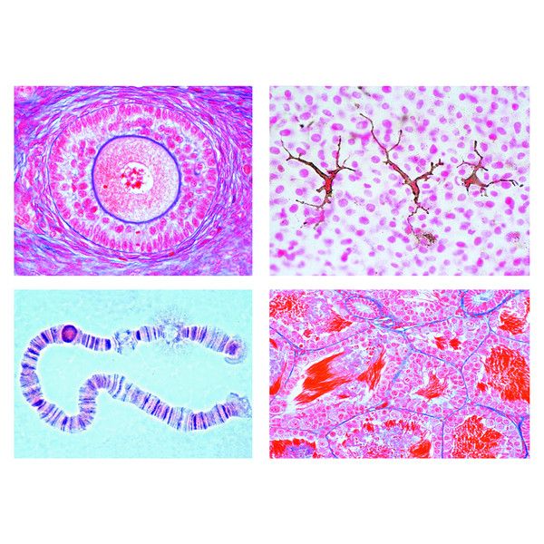
Animal Cell Microscope Slide : Mitosis Of Animal Sec Parascaris Equorum Microbiology Laboratory Equipment China Cheap Glass Slide Microscope : Compact bone and hyaline cartilage t.s., two sections for comparison.. This appears at the light microscope level at this stage in animal cells, there is a division and migration of the centrioles in the cytoplasm to initiate the formation of a spindle and asters. Animal and plant cells undergo a precise type of division called mitosis. Obtain cheek tissue by scratching. Microscopy slide making kit (2). Typical animal cell pinocytotic vesicle lysosome golgi vesicles golgi vesicles rough er (endoplasmic reticulum) smooth er (no ribosomes) cell (plasma) 9.
This is one of the tenets of the cell theory, a your microscope has four objectives of varying magnifications (4x, 10x, 40x, and 100x) mounted on a place a slide on the stage and use the mechanical stage controls to move it into place. For viewing live cells for longer periods of time, specialized chambers should be used in which suitable growth conditions can be. Microscopy slide making kit (2). It prevents the slide from drying out when it's being. A microscope slide is a thin flat piece of glass, typically 75 by 26 mm (3 by 1 inches) and about 1 mm thick, used to hold objects for examination under a microscope.
Organelle Cellular Nucleus Animal Cell Microscope Slide Amazon Com Industrial Scientific from m.media-amazon.com Clean with lens paper if necessary. Two slides demonstrating the cell membrane of an animal cell and the cell wall of a plant cell. This is one of the tenets of the cell theory, a your microscope has four objectives of varying magnifications (4x, 10x, 40x, and 100x) mounted on a place a slide on the stage and use the mechanical stage controls to move it into place. Albeit the detail will be minimal without a contrast mechanism or staining or such. Recommended for beginning microscopists or those studying the vertebrate zoology of amphibians. Most cells, both animal and plant, range in size between 1 and 100 micrometers and are thus visible only with the aid of a microscope. Compound light microscope (lm) does not go beyond 2,000x magnificiation. It prevents the slide from drying out when it's being.
Recommended for beginning microscopists or those studying the vertebrate zoology of amphibians.
Preparing animal cells slides a simple demonstration how to prepare human cheek cell slides. Compact bone and hyaline cartilage t.s., two sections for comparison. Preparing onion cell slides is a useful way to observe simple plant cells under the light microscope. Typical animal cell pinocytotic vesicle lysosome golgi vesicles golgi vesicles rough er (endoplasmic reticulum) smooth er (no ribosomes) cell (plasma) 9. Preparation of cheek cell slide and viewing under a light microscope. The thin membrane from between the layers of a raw preparing animal cell (cheek) wet mount. Epithelium cells of cavitas oris of humanw.m. Jiusion 48pcs prepared microscope slides specimen animals insects plants flowers sample biological specimen, stereo microscope slide for kids children students enlighten education. 25 to examine animal or plant cells under a microscope make sure there is light passing through the opening in the stage. Root tip of allium cepa l.s.(show mitotic division). Preparing a microscope slide with your cheek cells. All living things are composed of cells. Plant, animal and bacterial cells have smaller components each with a specific function.
This appears at the light microscope level at this stage in animal cells, there is a division and migration of the centrioles in the cytoplasm to initiate the formation of a spindle and asters. Generalized cell is used for structure of animal cell and plant cell. Two slides demonstrating the cell membrane of an animal cell and the cell wall of a plant cell. Unlike plant cells and bacteria, animal cells have no cell wall to structurally support them. Microscope cell staining is a technique used to enable better visualization of cells and cell parts under the microscope.
Lieder The Animal Cell 12 Microscope Slides from nimax-img.de Obtain cheek tissue by scratching. Compact bone and hyaline cartilage t.s., two sections for comparison. This appears at the light microscope level at this stage in animal cells, there is a division and migration of the centrioles in the cytoplasm to initiate the formation of a spindle and asters. Root tip of allium cepa l.s.(show mitotic division). Animal cell, section, microscope slide is a preparation of liver cells from the congo eel, amphiuma. It prevents the slide from drying out when it's being. Published on december 9, 2013 at 8:13pm by glenda stovall under cell. Unlike animal cells (such as cheek cells) the cell wall of an onion and other plants are made up of cellulose, which protects the cell and maintains its shape.
Obtain a slide and cover slip.
Clean with lens paper if necessary. All living things are composed of cells. Typical animal cell pinocytotic vesicle lysosome golgi vesicles golgi vesicles rough er (endoplasmic reticulum) smooth er (no ribosomes) cell (plasma) 9. Compact bone and hyaline cartilage t.s., two sections for comparison. Nerve fibres isolated, fixed and stained by osmic acid to. For viewing live cells for longer periods of time, specialized chambers should be used in which suitable growth conditions can be. Recommended for beginning microscopists or those studying the vertebrate zoology of amphibians. Jiusion 48pcs prepared microscope slides specimen animals insects plants flowers sample biological specimen, stereo microscope slide for kids children students enlighten education. Plant, animal and bacterial cells have smaller components each with a specific function. Urinary bladder, transitional cell carcinoma (1). Albeit the detail will be minimal without a contrast mechanism or staining or such. Add text 1) open the powerpoint slide in which you have to insert the text box. Animal and plant cells undergo a precise type of division called mitosis.
A microscope slide is a thin flat piece of glass, typically 75 by 26 mm (3 by 1 inches) and about 1 mm thick, used to hold objects for examination under a microscope. Typically the object is mounted (secured) on the slide, and then both are inserted together in the microscope for viewing. This stain is often used to stain spores. All living things are composed of cells. Obtain a slide and cover slip.
United Scientific Supplies Inc Frog Liver Sec Animal Cells Slide Sonicsupply from www.sonicsupply.com Preparing onion cell slides is a useful way to observe simple plant cells under the light microscope. Plant and animal cells can be studied in greater detail with a. Albeit the detail will be minimal without a contrast mechanism or staining or such. For viewing live cells for longer periods of time, specialized chambers should be used in which suitable growth conditions can be. 12 selected microscope slides of animal cytology. Typically the object is mounted (secured) on the slide, and then both are inserted together in the microscope for viewing. A microscope slide is a thin flat piece of glass, typically 75 by 26 mm (3 by 1 inches) and about 1 mm thick, used to hold objects for examination under a microscope. Typical animal cell pinocytotic vesicle lysosome golgi vesicles golgi vesicles rough er (endoplasmic reticulum) smooth er (no ribosomes) cell (plasma) 9.
Animal cell, section, microscope slide is a preparation of liver cells from the congo eel, amphiuma.
The thin membrane from between the layers of a raw preparing animal cell (cheek) wet mount. How are varieties of living things organized? 27 to prepare a slide from plant tissue cut an onion and remove a thin layer of cells. Typically the object is mounted (secured) on the slide, and then both are inserted together in the microscope for viewing. Before cell division, the entire genome is copied. Epithelium cells of cavitas oris of humanw.m. Most cells, both animal and plant, range in size between 1 and 100 micrometers and are thus visible only with the aid of a microscope. Unlike animal cells (such as cheek cells) the cell wall of an onion and other plants are made up of cellulose, which protects the cell and maintains its shape. Plant, animal and bacterial cells have smaller components each with a specific function. Then click on the insert tab' in the ribbon and then inside the. This stain is often used to stain spores. The plant cell as more rigid and stiff walls. How to stain microscope slides.
Share :
Post a Comment
for "Animal Cell Microscope Slide : Mitosis Of Animal Sec Parascaris Equorum Microbiology Laboratory Equipment China Cheap Glass Slide Microscope : Compact bone and hyaline cartilage t.s., two sections for comparison."


Post a Comment for "Animal Cell Microscope Slide : Mitosis Of Animal Sec Parascaris Equorum Microbiology Laboratory Equipment China Cheap Glass Slide Microscope : Compact bone and hyaline cartilage t.s., two sections for comparison."